This site is a work in progress. Please be patient.
BASIC TISSUE LOOKALIKES
Fried Eggs vs. Marshmallows
Simple Squamous Epithelium vs. Adipose Tissue
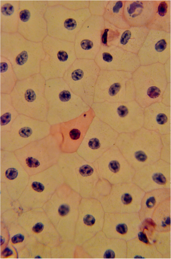
Simple Squamous Epithelium surface view
|
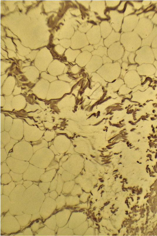
Adipose Tissue
|
Note the position fo the nuclei. They are central in this view of simple squamous epithelium, but peripheral in adipose tissue.
Cubes vs. Columns
Simple Cuboidal Epithelium vs Simple Columnar Epithelium
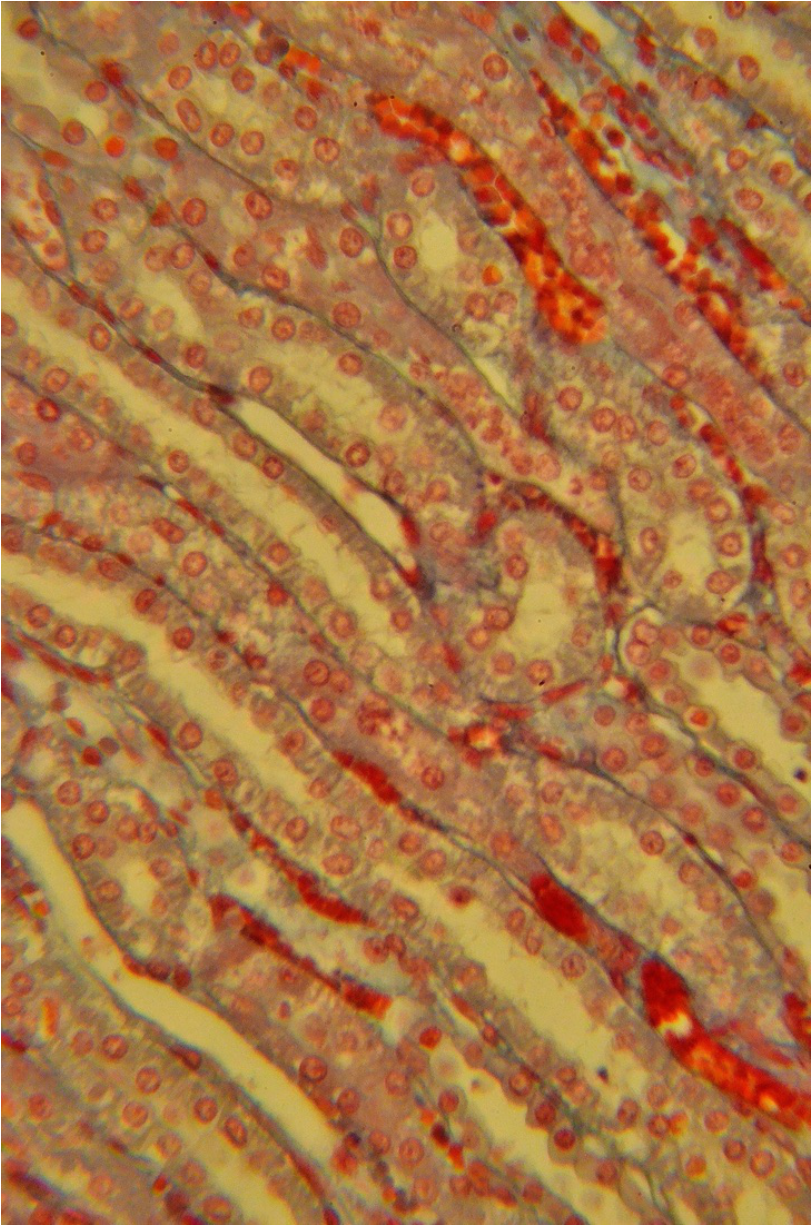
Simple Cuboidal Epiothelim
|
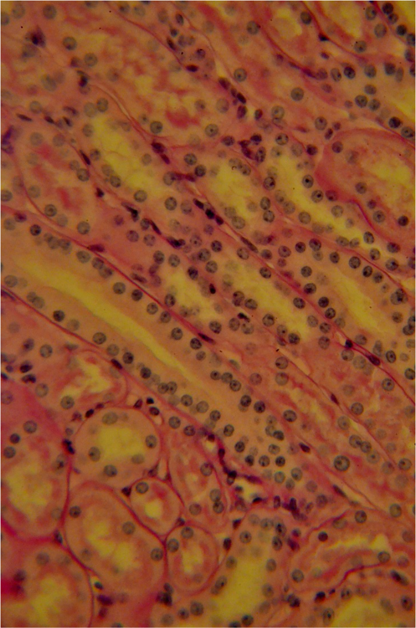
Simple Columnar Epithelium
|
Note that these are single layered tissues. Simple cuboidal epithelium is a principle tissues of non-vascular tubes. Simple Columnar Epithelium can also make tubes, but is more associated with linings. Note the position of the nuclei: In Simple Cuboidal Epithelium, the nucleus is central to the cell; in Simple Columnar Epithelium can be basal or apical, but rarely central. The image on the right shows a mixture of Simple Cuboidal and Simple Columnar Epithelia, Note too that the cytoplasm is also squarish in simple cuboidal epithelium and elongated in simple columnar epithelium, hence their names.
Tubular Stacks: Circles vs. Columns:
Stratified Cuboidal Epithelium vs. Stratified Columnar Epithelium
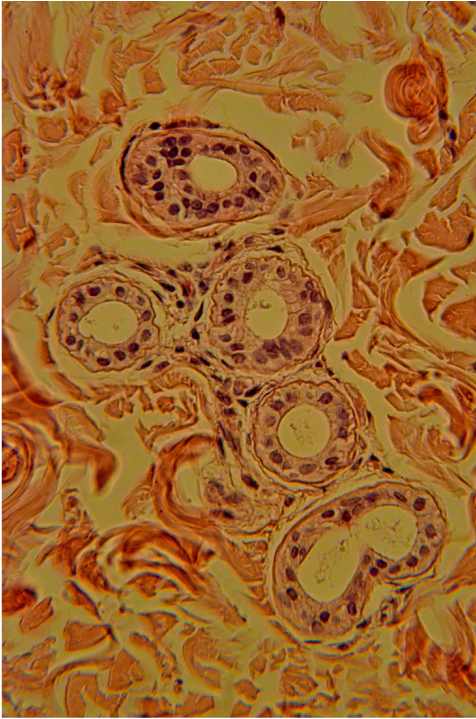
Stratified Cuboidal Epithelium
|
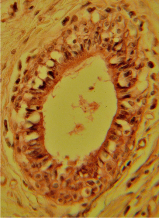
Stratified Columnar Epithelium
|
Of the two tissues, stratified cuboidal is the more common. Simple cuboidal often times appears as stacks of circles, rather than stacks of squares. Stratified cuboidal epithelium also makes the follicular and granulosa cells of the ovary. By contrast, column-shaped cells can always be detected in stratified columnar. I can't stress how rare this tissue is. I have seen it in salivary gland duct work, the eyelid, and the epiglottis.
Peacock Tail vs. Flattop
Transitional Epithelium vs. Stratified Squamous Epithelium, unkeratinized
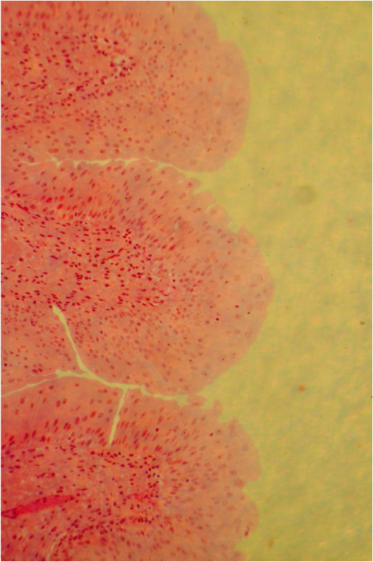
Transitional Epithelium
|
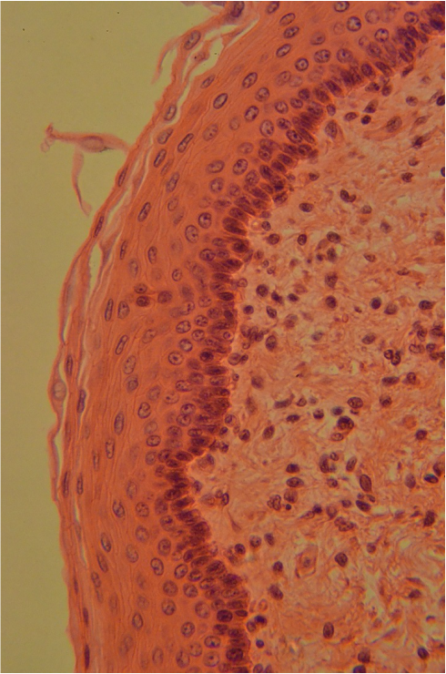
Stratified Squamous Epithelium, unkeratinized
|
Both Transitional Epithelium and Stratified Squamous Epithelium are mulch-layered tissues. Transitional resembles a Peacock's tail or water coming from as sprinkler. The surface tissue is billowy, often with nuclei at the apical (free) end. Stratified squamous, epithelium is always flat at the apical end. These tissues get harder to tell apart in distended Transnational epithelium, although here, the cells are more uniformly flat, while the flatness is restricted to the apical surface cells in Stratified Squamous Epithelium, unkertinized.
Columns vs. Stack Wannabes
Simple Columnar Epithelium vs. Psuedostratified Epithelium
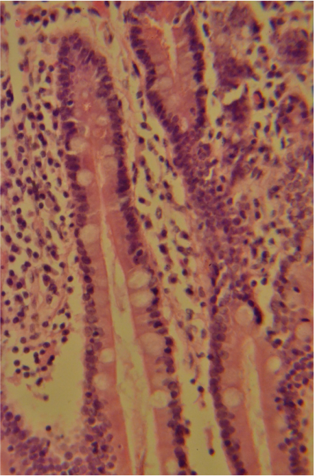
Simple Columnar Epithelium
|
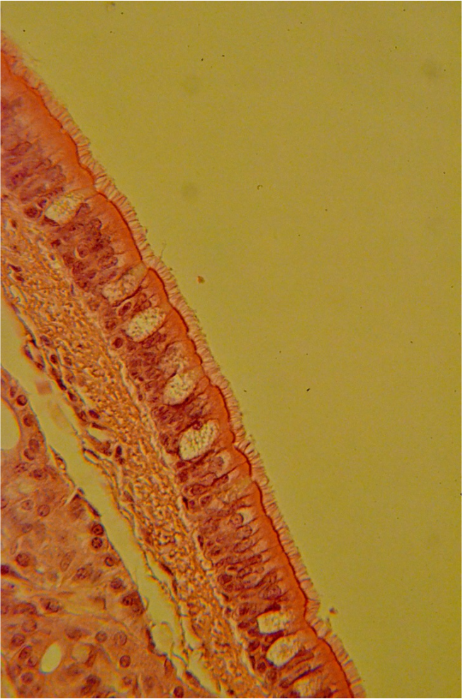
Pseudostratified Epithelium
|
Simple Columnar Epithelium is often confused with Psuedostratified Epithelium because slide prep for the Simple Columnar Epihtleium is often thick. As a result, multiple nuclei appear in this issue, giving it a stratified appearance. Pseudostratied Epithelium can be columnar or cuboidal in shape, but in most cases it is ciliated. There a a few cases of ciliated simple columnar epithelium, but the nuclei appear much more uniform in arrangement, even if multiple layers of cells are present. Microvili can also be found in simple columnar epithelium which resemble cilia, but these appear think and less brush-like. Simple Columnar Epithelium is associated primarily with the alimentary canal, fallopian tubes, and certain glands. Pseudostratfied epithelium predominates the repository system, although ti can also be found in the male reproductive systeml.
Flattops: Flaky vs. Smooth
Stratified Squamous Epithelium: keratinized vs. unkeratinized
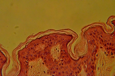
Stratified Squamous Epithelium , kertinized
|
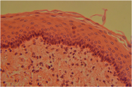
Stratified Squamous Epithelium, unkertinized
|
Both stratified squamous tissues earn their name from the squamous cells on the apical (free) surface. The term squamous means scale, and when viewed from the side, the cells appear flat. Dead cells of the keratinized form are devoid of nuclel and often appear as thin crusty layers. The aplcal cells of the unkeratinzed form contain obvious nuclei. The cells below appear cuboidal or even columnar. Nobody cares, since its the apical surface cells that the tissues are named for.
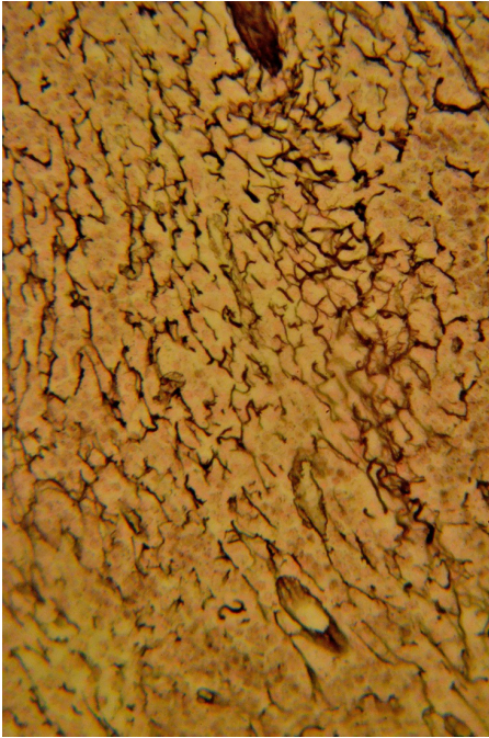
Fish Nets & Spider Webs
Reticular Tissue vs. Areolar Tissue vs. Elastic Tissue, regular form
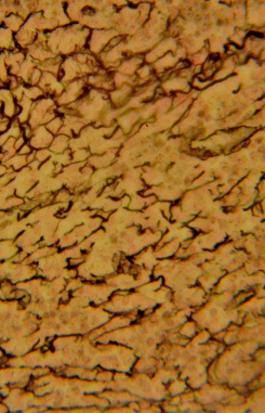
Reticular Tissue
|
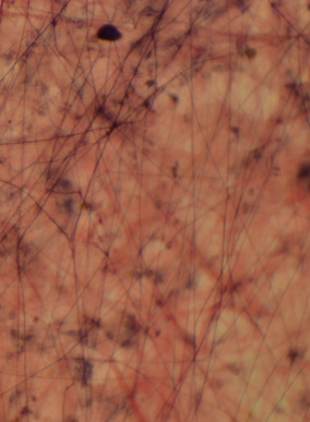
Areolar Connective Tisse
|
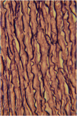
Elastic Connective Tissue, regular form, stained to show elastic fibers
|
Dark fibers appear in all three of these tissues, giving them all a net-like appearance. Reticular tissue fibers are made of type III collagen and are best seen magnified at 40x or more. They are delicate and only around 1 mm in length. You will wind easily them in the lymph nodes, and depending upon the stain, they may appear in the spleen are liver. Areolar connective tissue has long dark fibers interlaced with thick pink collagen. Fibroblasts, macrophages, mast cells and various white blood cells take up residence here, resembling flies caught in a spider web. The irregular form of elastic tissue is very uncommon has shorter fibers. By contrast, the regular form, (above) has long fibers and a robust appearance. They are also typically more wavy than the elastic found in areolar connective tissue.
Squiggles vs Nets
Elastic Connective Tissue, irregular form vs Reticular Tissue
|
|
Strips
Elastic Tissue, regular form vs. Dense Fibrous Tissue, regular form vs. Bone
The irregular form of elastic tissue requires special stain to see well. Here we see it as dark amber wavy lines (~80x). By contrast, Reticular tissue has much thinner and shorter fibers, most obvious in the lymph nodes (40x). Again, with proper staining it can be seen in the spleen and liver.
Elastic Connective Tissue, Regiular Form
|
Dense Fibrous Connective Tissue, irregular form
|
Decalcified Bone
|
The regular form or Elastic Tissue appears as long noodles, with or without nuclei present. By contrast dense irregular connective tissue is chucky and haphazard in appearance. Bone is smooth with a complete matrix.,
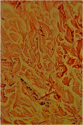
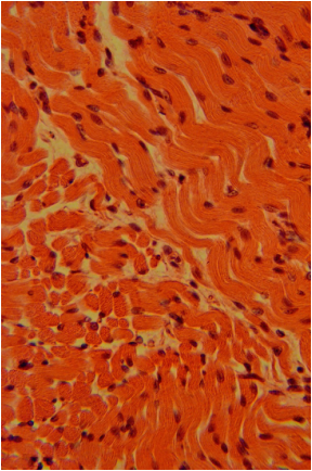
Highways vs. Fish Schools vs. Chunks
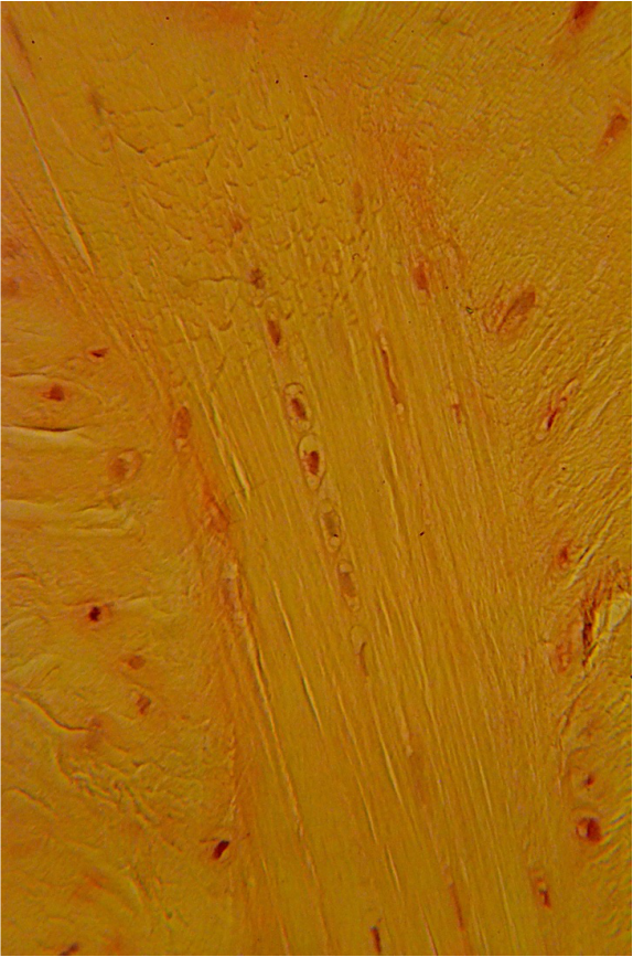
Dense Fibrous Tissue, regular form vs. Smooth Muscle vs. Elastic tissue, regular form
Dense Firbous Connective Tissue, regular form
|
Smooth Muscle
|
Elastic Connective Tissue regular Form
|
Dense Fibrous connective Tissue resembles a multi-lane highway; the nuclei are always linearly arranged. Its matrix is more or less a continuous band. Smooth muscle cells can often be distinguished form one another, having no matrix. Smooth muscle often have a school of fish appearance, especially when the cut is irregular. The nuclei never follow a truly linear pattern. Elastic tissue regular is most similar to dense fibrous connective tissue regular form, but it's matrix is separated into distinct ribbons.
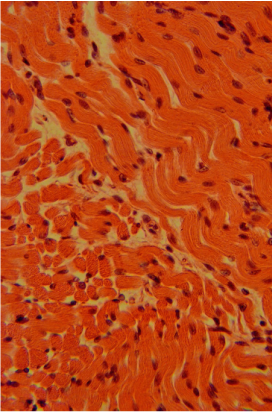
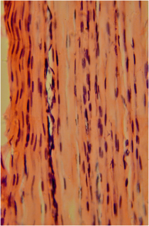
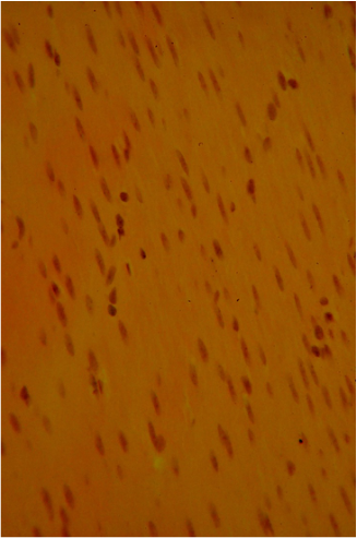
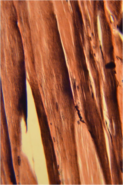
Banded Tissues (Striations)
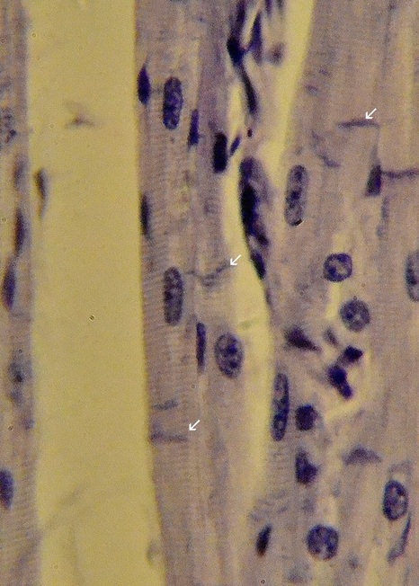
Skeletal Muscle vs. Cardiac Muscle
|
|
Both cardiac and skeletal muscle have visible striations (dark and light bands) and are the only tissue to posses these. They are due to the presence of sarcomeres--actin and myosin protein arrangements that form the tissue's cytoskeleton. Cardiac striations are always much fainter that skeletal muscle. The image above is magnified to approximately 80x, roughly twice that of the adjacent skeletal muscle (40x) to make the striations more visible. Skeletal muscle cells always mult-inucleated although nuclei are not always visible as in our image. Cardiac muscle cells have single nuclei or sometimes double. One of the best distinguishing features is the the presence of intercalated disks in cardiac muscle (see arrows). These appear as extra dark striations scattered among the faint ones. All striations in Skeletal muscle will be uniform.
Frog Eggs
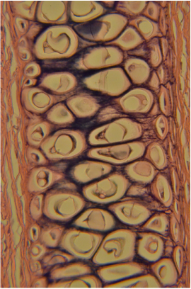
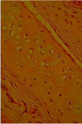
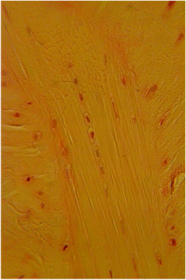
Elastic Cartilage vs. Hyaline Cartilage vs. Fibrocartilage
Elastic Cartilage
|
Hyaline Cartilage
|
Fibrocartilage
|
In all three cases, cartilage cells are housed in lacunae, making them appear egg-like. The matrix of elastic cartilage has elastic fibers within it, while Hyaline cartilage as a smooth matrix. Fibrocartilage has matrix that often appears to have small breaks in it, these breaks can resemble fibers. The chondrocytes of elastic and hyaline cartilage are randomly dispersed,while the chondrocytes of fibrocartilage are often linear, particularly in older tissue. New growth of fibrocartilage often contains multiple chondrocytes within a single lacuna. As this tissue ages, they develop a linearity which continues as newly formed matrix separates them to individual lacunae.
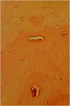
Holes and Sheets
Fibrocartilage vs. Bone
Fibrocartilage
|
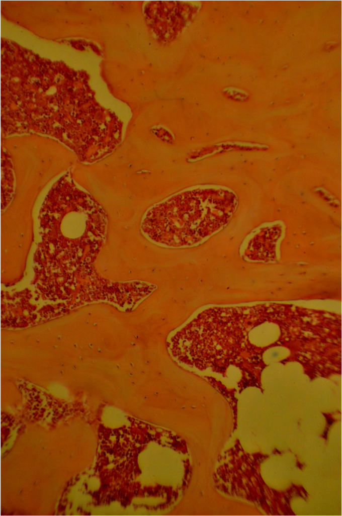
Decalcified Bone
|
Chondrocytes of cartilage and osteocytes of bone are both housed in lacunae, often making these tissues look similar. This i particularly true with fibrocartilage and woven bone. Fibrocartilage is restricted to the symphysis pubis and will not have large holes within it. These holes in are created by osteoclasts that remodel bone and the holes usually contain red bone marrow.
Bone Again!
Compact Bone: Transverse vs. Longitudinal sections
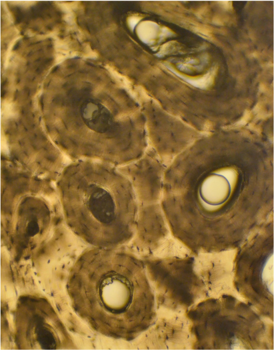
Compact Bone: Transverse Section
|
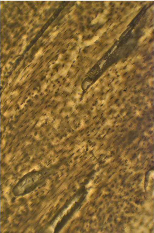
Compact Bone: Longitudinal section
|
No other tissue resembles compact bone. Note the osteons on the left, which resemble tree trunks. The black spots in both sections are bare lacunae where oestocytes once resided. This ground bone preparations removes all living cells but keeps their lacunae. The lacunae and the spidery canaliculi that project form them, reveal the shape and location of the osteocytes (See Connective Tissue: Bone).
Althogther Now!
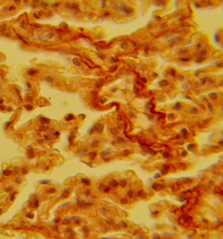
Hylaline Cartilage vs. Areolar Connective Tissue vs. Smooth Muscle
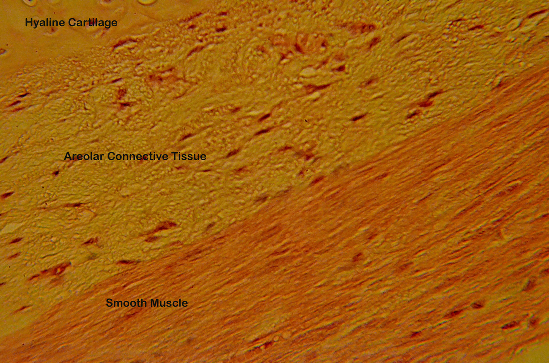
The image above is a 40x view of a region of the trachea. Note the texture and cellular differences between the three featured tissues. Hyaline cartilage has a fish egg appearance. Areolar connective tissue has a loose appearance while smooth muscle looks more dense. Can you see the directional change in muscle cell arrangement? The muscle cells are longitudinally cut in the region adjacent to the areolar connective tissue and more transverse cut at the lower region of this tissue.
Guess the Ganglion
Sympathetic vs. Dorsal Root Ganglia
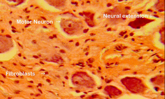
Sympathetic (autonomic) Ganglion
|
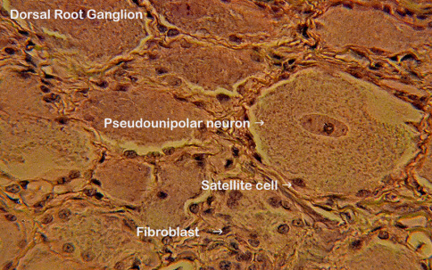
Dorsal Root Ganglion
|
Ganglia can be confusing. Note the central nuclei of the psuedounipolar cells of the dorsal root ganglion vs off center nuclei of the sympathetic chain ganglia. Typically satellite cells will be abundant in both.
